Multimodal computer-aided diagnosis system for adolescent mental health
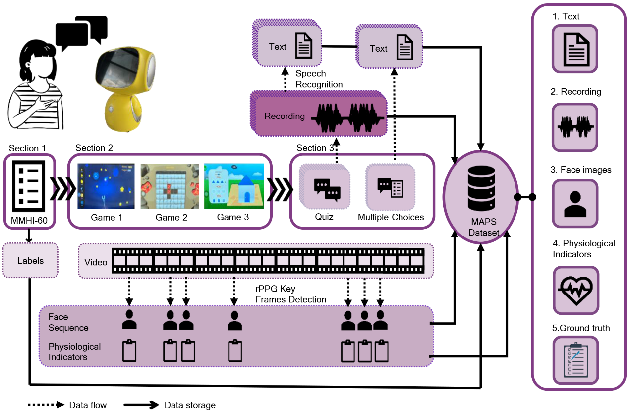
The timely identification of mental disorders in adolescents is a global public health challenge. We have developed a humanoid robot equipped with well-designed emotional stimuli that facilitates the acquisition of the Multimodal Adolescent Psychological Screening (MAPS) dataset, including facial images, physiological indicators, audio recordings, and textual transcripts. Based on this, we propose GAME (Generalized model with Attention and Multimodal EmbraceNet, a generalized model based on distance-weighted attention mechanisms and multimodal feature fusion for adolescent mental disorders screening. In the future, we will continue to explore the development of more comprehensive interactive robots and personalized human-computer interaction methods, as well as powerful and interpretable multimodal learning networks. At the same time, we continue to pay attention to the challenges of missing modality data and unbalanced data distribution in practical application scenarios.
Nucleic acids detection with CRISPR/Cas systems
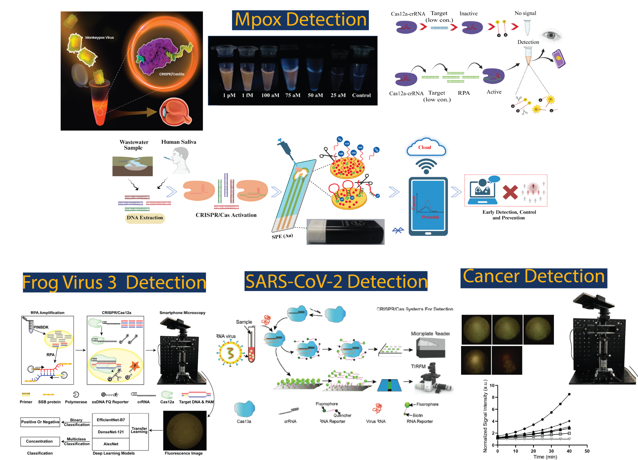
Since 2017, our team has developed CRISPR/Cas-based integrated sensing systems tailored for the detection of RNA and DNA viruses. Leveraging the collateral activities of Cas13a and Cas12a, we have successfully developed CRISPR diagnostics for detecting Ebola Virus RNA (Qin et al., ACS Sens., 2018), SARS-CoV-2 RNA (Qian et al., Microchim. Acta., 2024), African Swine Fever Virus DNA (Qian et al., Biosens. Bioelectron., 2020), Mpox Virus DNA (Chen et al., J. Med. Virol., 2022), and Frog Virus 3 DNA (Lei et al., ACS Omega, 2023), achieving sensitivities ranging from femtomolar to attomolar levels. Currently, we are developing next-generation multiplex theranostic platforms to combat bacterial, viral, and fungal infections.
IMAGING GENOMIC Loci with cas9
Imaging chromatin dynamics is crucial to understand genome organization and its role in transcriptional regulation. Recently, the RNA-guidable feature of CRlSPR-Cas9 has been utilized for imaging of chromatin within live cells. However, these methods are mostly applicable to highly repetitive regions, whereas imaging regions with low or no repeats remains as a challenge. To address this challenge, our lab has proposed two strategies:
(1) Utilize single-guide RNAs (sgRNAs) integrated with up to 16 MS2 binding motifs to enable robust fluorescent signal amplification (Left figure). These engineered sqRNAs enable multicolor labeling of low-repeat containing regions using a single sgRNA and of nonrepetitive regions with as few as four unique sgRNAs.
(2) Leveraging off-target binding to enhance the imaging efficiency at low-repetitive genomic loci (Right figure). We found that there are ten thousand of sites with more than 20 off-target binding sites located within a 10 kb vicinity of their target sites (colocalized off-target repeats) in the human genome. By strategically designing the sgRNA to target the genomic loci with colocalized off-target repeats, the imaging efficiency of low-repetitive genomic loci can be significantly improved.
Using these tools, we can track the locations of native chromatin loci throughout the cell cycle and determine differential positioning of transcriptionally active and inactive regions in the nucleus.

Application of Computational methods in biomedicine and public health
Since 2019, we start the interdisciplinary research on the development of computational method and its application in biomedicine and public health. We have built collaboration with many domestic hospitals to develop AI assisted diagnosis method with ethical approval. The ongoing projects include: (1) MRI lesion segmentation for acute cerebral infarction, (2) spinal and brain tumors segmentation of MRI, (3) tongue diagnosis in Traditional Chinese Medicine (TCM), (4) Multimodal integration for adolescent mental health screening, (5) Creation of multimodal atlas of police Canine brain.

Development of biomedical robot
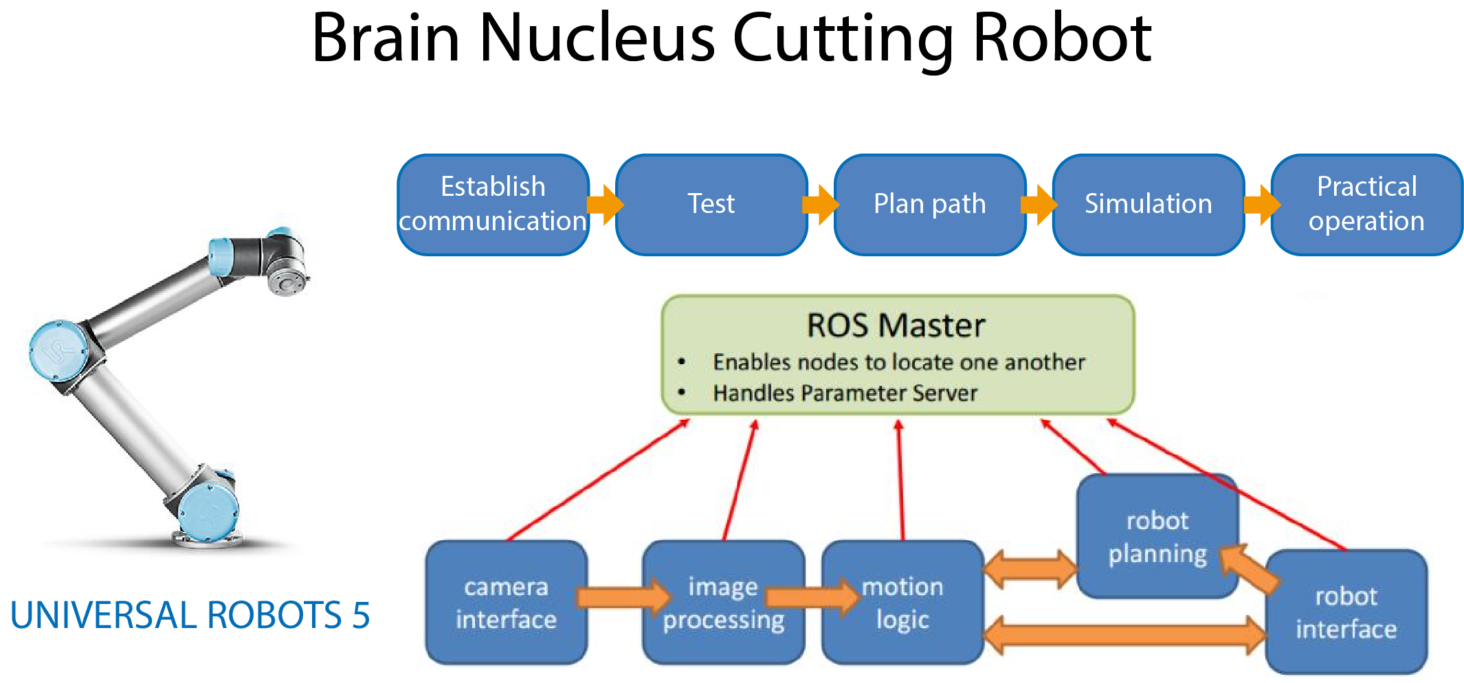
Our brain is organized into multiple correlated functional domains. Our initial goal is dissect individual functional domains with path-design robotic arm. To extend the function of brain domain segmentation, we will develop imaging guided surgical robot. We use multi-axis mechanical arm to realize the automation in biomaterial applications. The current applications include segmentation of irregular brain nuclei, preparation of ultrathin agarose gel, and acquisition of patients’ pupil image during surgery, etc.
Flexible ionic conductive elastomers for human machine interaction and healthcare
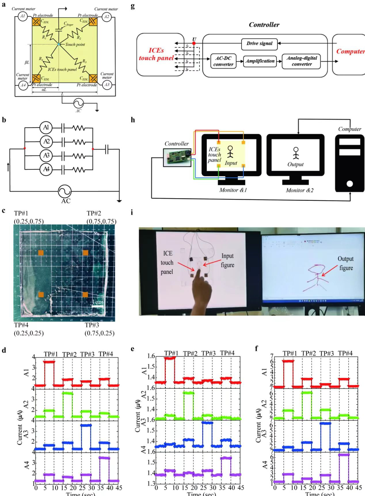
1) Stretchable transparent trackpad
Stretchable transparent trackpads have received a lot of research attention, in which ionic conductors of hydrogels/ionogels play a key role. However, hydrogels/ionogels often have inherent limitations. These devices are affected by dehydration or evaporation of liquid solvents at high temperatures, or prolonged exposure to the open air, which can make their ion conductivity and stretch worse. Hydrogels dry out easily in actual use, leading to equipment failure. Hydrogels freeze at low temperatures, resulting in a loss of stretchability and self-healing ability, and a significant decrease in ionic conductivity. Several strategies have been proposed to address the above problems, such as the introduction of chemicals or salts in hydrogels, which can delay dehydration and lower the freezing point, but cannot completely prevent dehydration. Hydrogel equipment requires the use of hydrophobic materials for sealing and coating to effectively retain moisture, however, the strong tack of hydrogels/elastomers requires complex procedures. While ionic gels have recently received a great deal of attention due to their non-volatilization, good thermal stability, and ionic conductivity, the challenge is liquid leakage when deformed. Therefore, the ideal stretchable touchpad would utilize elastomers as electrified materials and electrodes (Chemical Engineering Journal, 2022, 451, 138672)。
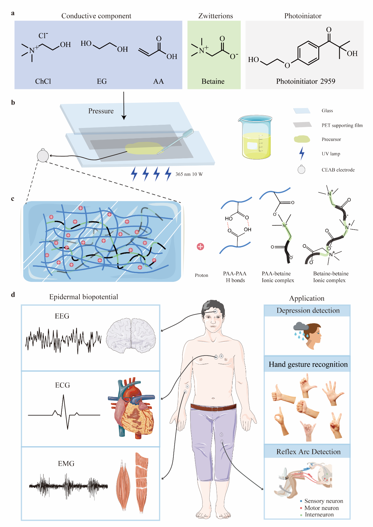
2) Skin-like electrodes for epidermal electrophysiological acquisition, hand gesture recognition and depression detection
Flexible ionic conductive electrodes, as a fundamental component for electrical signal transmission, play a crucial role in skin-surface electronic devices. Developing a skin-seamlessly electrode that can effectively capture long-term, artifacts-free, and high-quality electrophysiological signals, remains a challenge. Herein, we report an ultra-thin and dry electrode consisting of deep eutectic solvent (DES) and zwitterions (CEAB). Compared to standard Ag/AgCl gel electrodes, our electrodes exhibit significantly lower reactance and noise in both static and dynamic monitoring. This improvement can be attributed to skin-like mechanical properties, outstanding adhesion, and high electrical conductivity. Consequently, they excel in consistently capturing high-quality epidermal biopotential signals, such as the electrocardiogram (ECG), electromyogram (EMG), and electroencephalogram (EEG) signals. Furthermore, we exhibit the promising potential of the electrode in clinical applications by effectively distinguishing aberrant EEG signals associated with depressive patients. Meanwhile, through the integration of CEAB electrodes with digital processing and advanced algorithms, valid gesture control of artificial limbs based on EMG signals is achieved, highlighting its capacity to significantly enhance human-machine interaction.
High quality fluorescence imaging systems
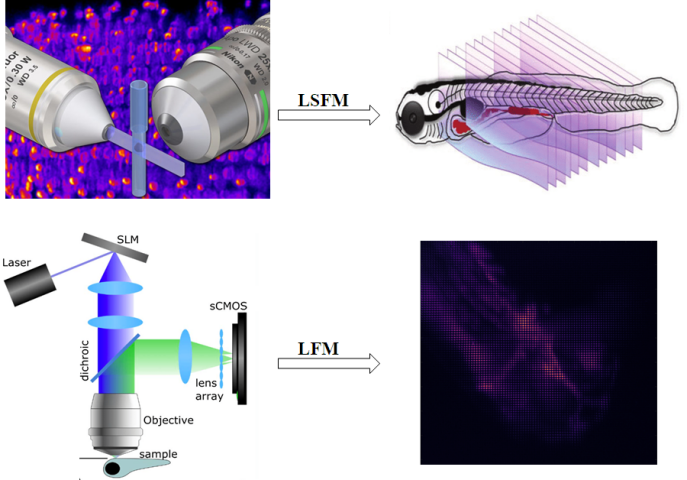
Light sheet fluorescence microscopy (LSFM) functions as a non-destructive tool to optically section and image tissues with subcellular resolution with a plane illumination. Since samples are exposed to a thin plane of light, photobleaching and phototoxicity are minimized compared to wide-field fluorescence, confocal or multiphoton microscopy. Light field microscopy (LFM) provides an elegant compact solution for scan-less volumetric imaging. It records rapid biological activities by taking a snapshot. Then a 3D scene of these activities can be recovered by post-processing the light field information. We are integrating the LSFM and LFM systems for the biomedical applications, such as the recording of neuronal activities, visualization of cardiac dynamics in small animals and blood flow imaging at single cell resolution. We are also committed to optimizing LFM-related image processing algorithms to realize high resolution and high throughput biomedical 3D imaging.
